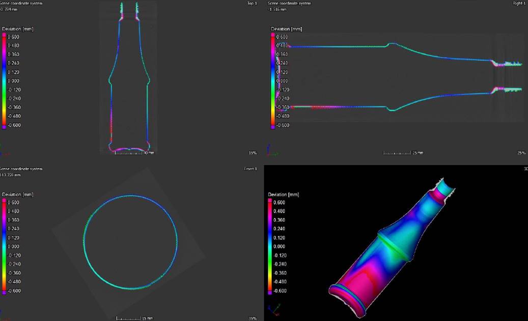
dimensional and wall thickness measurements
PRODUCTS
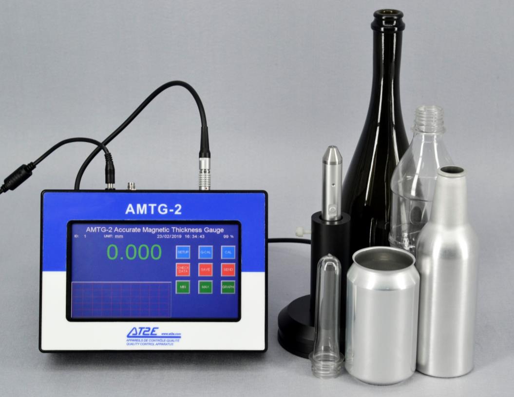
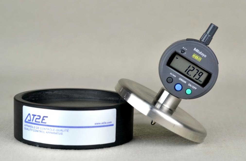
Accurate Magnetic Thickness Gauge
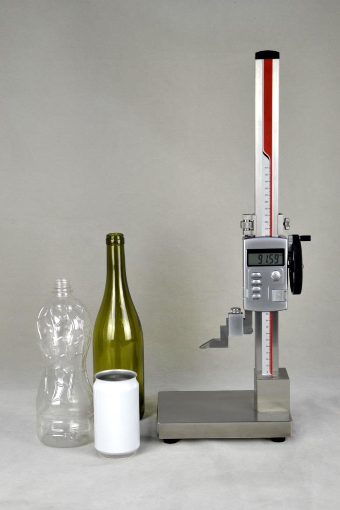
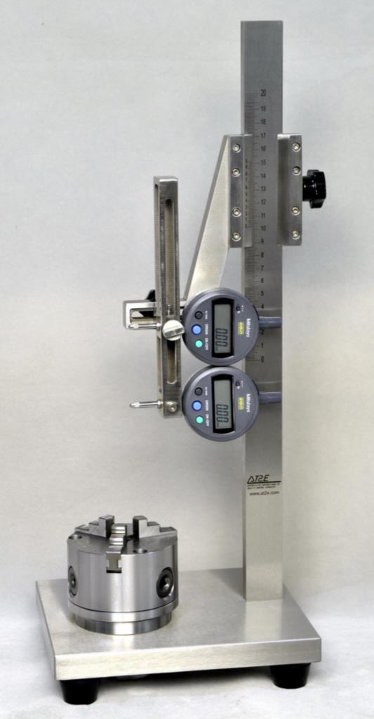
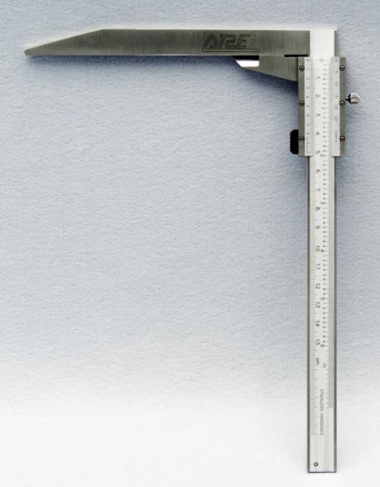
Height Gauge
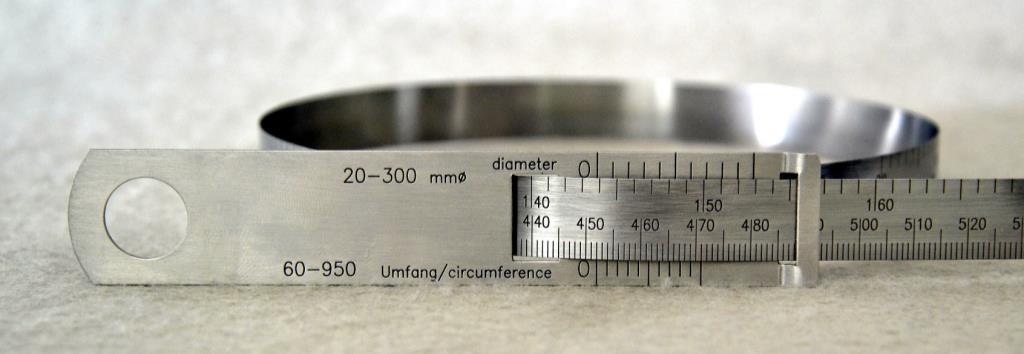
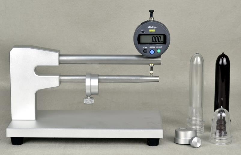
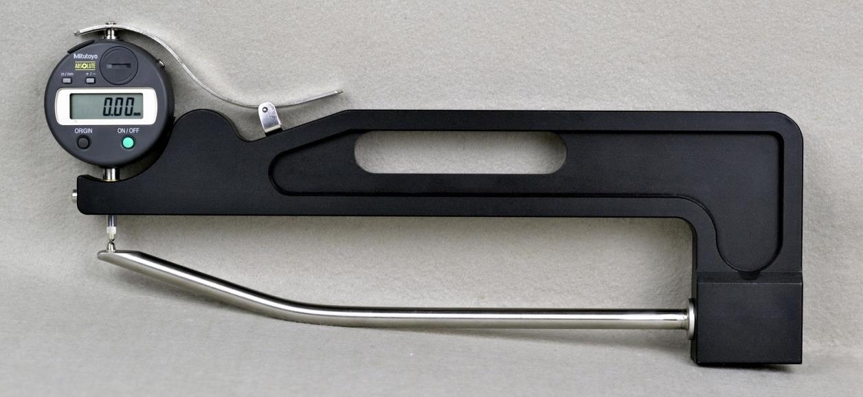
Circometer and Caliper
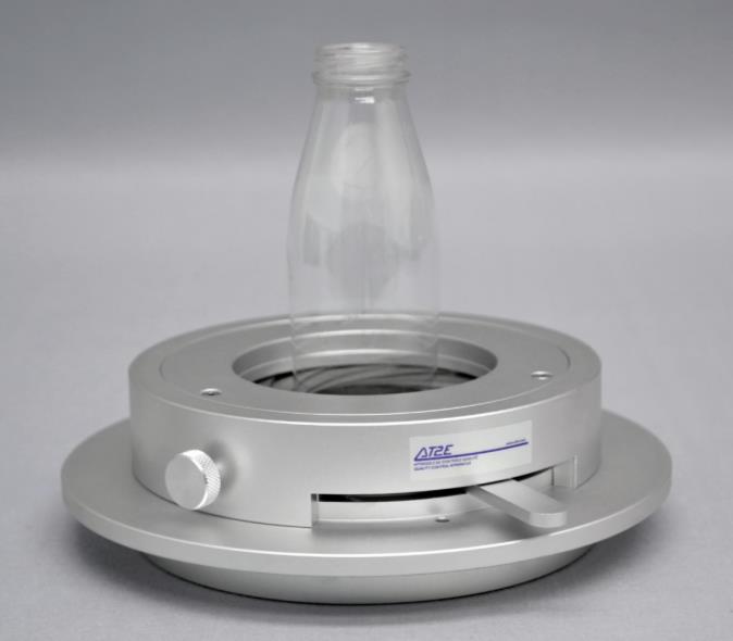
Base Clearance Gauge
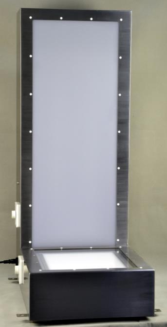
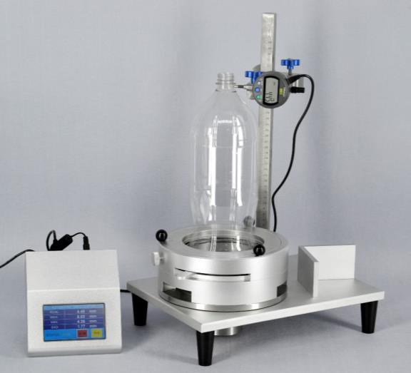
Glass Bottle Lightening System
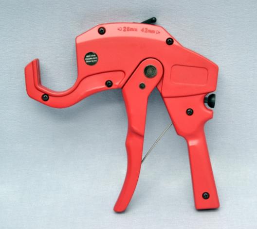
Preform Cutter
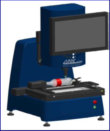
Preform & Bottle Measuring System
Perpendiculary Gauges
Thickness Gauges
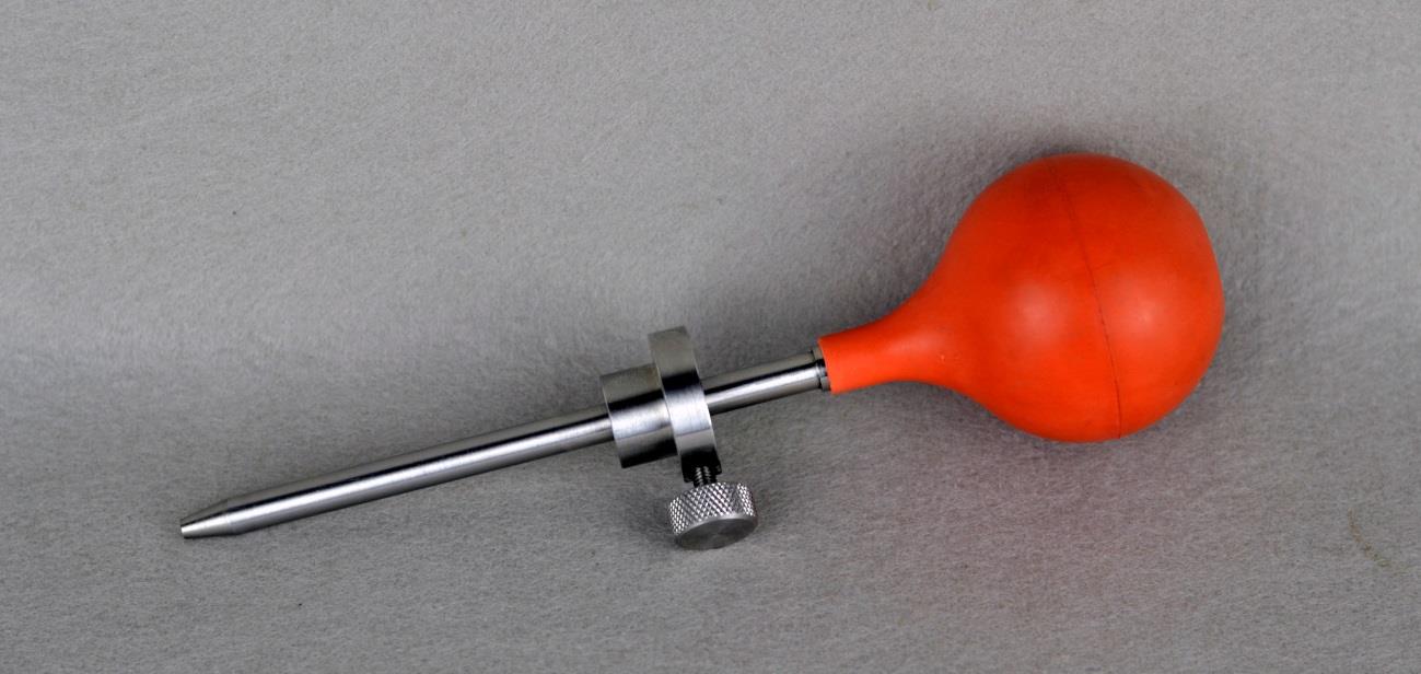
Fill Hight Gauges, Fill Hight Syringe
Automated Preform & Bottle Dimension Tester
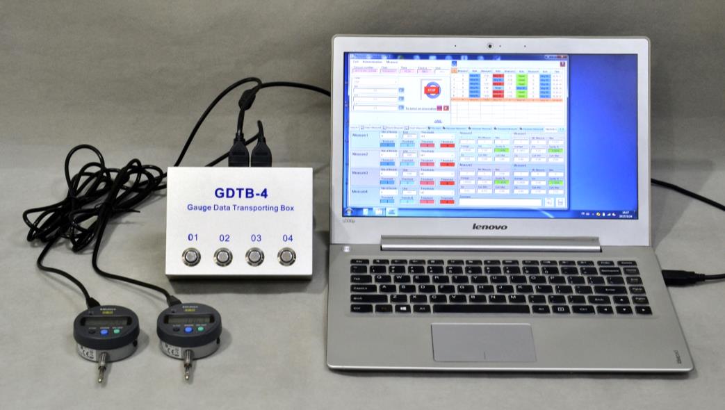
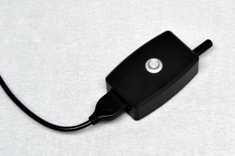
Gauge Data Transportation Box
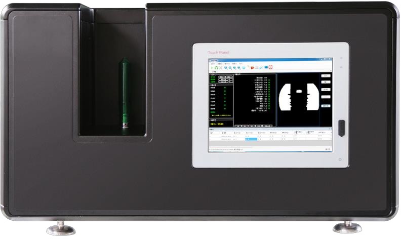
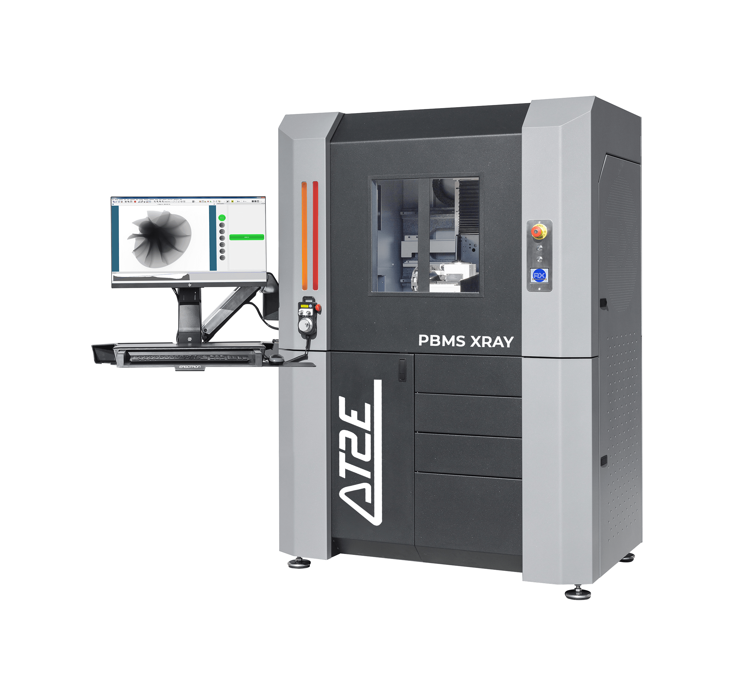
PBMS-XRay Preform& Bottle Measuring System X-RAY
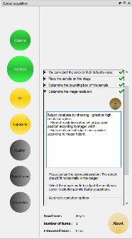
Description
Computed tomography system with an internal volume of 591 mm (W) x
685 mm (H) x 445 mm (D), with automatic door, equipped with a sealed X-ray generator (maintenance free), 130
kV, 5µm minimum focal spot size and max power 39 W, a flat panel detector with “real time” acquisition, high
precision mechanical rotationstage & 3 motorized axis, acquisition& reconstruction computer and
proprietary software to control the equipment and to reconstruct data.
Consists of
following configuration
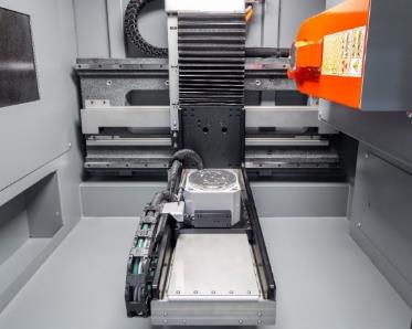
Lead protected cabinet with a large inside volume of:
445mm (W) x 591 mm (Z) x 685 mm (H)Motorized secure access door (automatic interlock) with large leaded
glass windows
PC control unit, Windows X-Act software license to drive
the equipment. Plugins including CT acquisition, live videos recording, dimensional measurements in
2D radio mode, learning macro mode, automation.
Unlimited X-Act viewer licenses that may be
redistributed or installed on an unlimited number of PCs in order toview radiographic images and
reconstructed tomographic volumes. Specific workstation with required specs for tomographic
reconstruction on GPU hardware parallelization cards (Windows 64 bits, 64 GB RAM, high performance
Graphic Card, Powerful processor Intel Xeon W-2125, x2 SSD (Raid 1) x2 hard drives of 6 TB mirrored
(Raid 1). This workstation will serve besides reconstruction as post-processing unit using e.g.
VGStudioMax, Avizo...
Dongle license for AT2E X-Act software CT reconstruction
module.
CE marking, compliant to NF C74-100 &17-DC-0591 radiation safety
standards
12 months’ warranty. Warranty includes AT2Esoftware maintenance and remote
support.
Global
Electrical Power: 230 V, 50 Hz, 3
kW
Room temperature for metrology use: active temperature control: +15 °C to +25 °C
Hygrometry:
less than 80 % relative humidity non-condensing
Maximum resolution: around 5 µm / voxel with a9 mm
diameter sample placed at 11mm from the output window (compatible with a temperature cell), 10 µm /
voxel for an 18 mm diameter sample.
True resolution: ability to visualize separately pair of lines on
a reconstructed tomographic slice: 5 µm
Maximum scanned volume: diameter180 mm - height 380
mm
Cabinet
Outer cabinet dimensions : 1325 mm
(W) x 890 mm (D) x 1865 mm (H)
Inner cabinet dimensions : 710 mm (W) x 604 mm (D) x 835 mm (H).
Maximum
height between rotation stage and cabinet ceiling: 685 mm (may vary depending on imager)
Maximum
distance generator/detector: approx. 590 mm
Focal spot to imager distance: 620 mm
Self-protected
cabinet with outside leak <0.5 µSv/h
Automatic door + interlock
One through port access for
user
Motorized sliding doors with large leaded windows to see inside unit.
Weight of the
equipment: 1020 kg
Mechanics
3 precision granite linear
motorized axis are used in this system and one precision rotation stage
- vertical shift: 275 mm
- lateral shift: 195 mm
- magnification: 480 mm
Motorized axes:
The mechanical system includes 3
precision granite motorized axes + 1 precision rotation stage:
Adjustment of the magnification by
moving therotation stage closer or further from the imager or generator
Axis 1: lateral axis for the
sample stage (190mm).
Axis 2: vertical axis for the sample stage (270mm).
Axis 3: zoom axis
mounted on granite-base (approx. 466 mm).
Maximum load on the rotation stage: 5 kg.
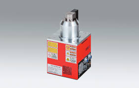
X-RayGenerator 130 kV
Sealed micro-focus
X-Ray generator with maximum voltage: 40 to 130 kV, 300 µA, maximum power: 39W and minimum focal spot
size: 5 microns.
Flat panel
High resolution digital
“real-time” flatpanel (25 x 32 cm active area, 2048 x 2560 pixels, size:124 µm).
2048Pixels x 2560 Pixels
Pixel size: 124 µm
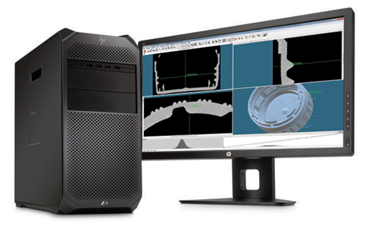
Computers
Acquisition computer
HP
Z4 G4 8 GB RAM, 2 x 256 GB SSD (Raid1)
Flat panel wide screen 27 inches, keyboard and mouse, DVD,
USB3 ports, Windows 10 64 bits.
Reconstructioncomputer (or equivalent)
HP Z4
G4 64 GB RAM, Intel Xeon W-2125 processor, Powerful graphic card Nvidia GF RTX 2080 (GPU), 2 x 256 GBSSD
(Raid 1) & 2 x 6 TB Hard drive (Raid1), Network card 1 Gb.
Flat panel wide screen 27 inches,
keyboard and mouse, USB3 ports, Windows 10 64 bits.
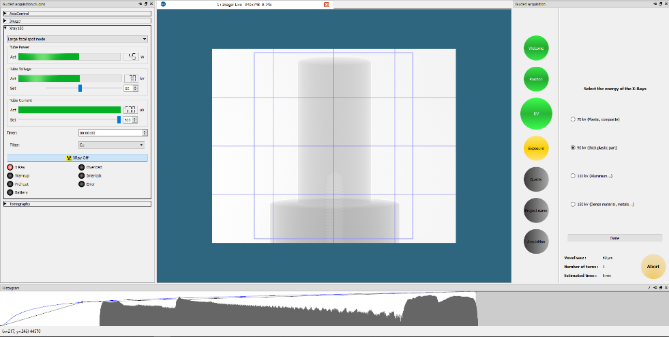
Software
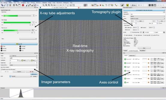
RXSolutions software
X-Act
Acquisition and CT system control. User has full control on hardware from AT2E X-Act.
X-ray tubes: Adjustable settings: voltage, current, focal mode (ifany). Automatic X-ray spot
size adjustment according to the voxel size
Imagers: Available settings (depending on imager):
binning and gainmodes, ROI, frame rate, black and gain calibration (includingmulti-gain)
Axes:
list of axes with individual control possible. Movement via GUIor handwheel. Absolute or relative
position, speed control, homing
Overlay: overlay tools to draw geometric primitives, measure
3Ddistances and angles, load and display STL file
Image processing: image to image mathematic
operation, 3D volum evisualization, STL extraction based on grey level threshold, MTF measurements
Execution of Automated sequences via macro / scripting
Load
acquisition and reconstruction profiles for sample series
Automatic calibration procedure of the
geometry of the system
CT acquisition
The CT acquisition plugin allows
tomographic acquisition with the following options:
Standard single rotation, Horizontal
stitching (“shift”),Vertical stitching (“stack”), Helical, Continuous rotation (local tomography, fast
acquisitions)
Reference acquisition for X-ray/sample movement compensation
Multiple
turns (acquisition split in several rotations for fast preview), Limited angle rotation, Laminography
(no rotation: planar3D acquisition)
Anti-ring shift
Automatic black and gain calibration
Stabilization sleep time
Automatic registration of the acquisition settings
Automation:
one-click acquisition and reconstruction start for series of samples
Real-time 2D video
radiographies
Guided acquisition mode for beginners and advanced user interface
In-situ and customization:
Ready to use serial port
communication plugin in X-Act software to control user hardware
“SDK” like package to implement your
own plugins (custom demand)
Java-like scripting language for scripts and console
CTreconstruction
The
reconstruction solution provided is a licensed plugin of X-Actsoftware called UniCT. It is a GPU
accelerated CT reconstruction module mainly based on filtered back-projection algorithm. A complete set
of tools is available to reconstruct acquisitions performed on PBMS XRAY system including:
Preview
reconstruction of a running acquisition
Any direction slice preview
Full geometry correction,
including automatic correction of therotation center
Focal spot drift, rings, bad pixel, beam
hardening, phase contrast, metal artefacts real time correction (slice preview)
Easy and intuitive 3D
optimization of the reconstructed volume: size, orientation and position of the bounding box. Possible
import of previous acquisition settings to allow direct co-registration of theslices (valid if the
sample was not removed from the stage or was placed at the same position)
Import menu to reconstruct
data from 3rd party CT systems (including parallel beam projections like synchrotron
acquisition)
TIF 16-bits image formats to allow user specific image processing before and/or after
reconstruction.
Automation: enqueue preset projects and run batch reconstructions.
Licensefree
X-Act main features
X-Act software can be used without any licenses and shared
between several users.
Load and display images or series of images in stack or in 3D.
Change
contrasts, colors, apply filters (contrast enhancement, denoising, visual effects…)
Mathematical
operations on images or image stack (subtract, mean, add…)
Merging or superimposition of images
Image
analysis tools: MTF measurement plot, grey scale profile
Extract and export point clouds (STL)
2D
and 3D measurements
Drawing tools using overlays
High quality video recording starting from image
stack
Conversion and compression of images (from 16-bits Tiff format to compressed format 8-bit Tiff,
jpg, png)
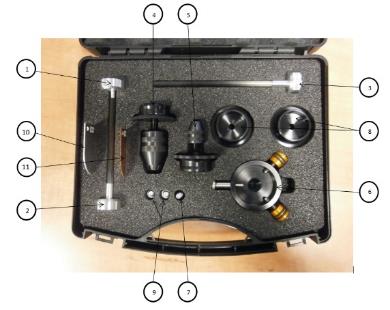
Accessorycase
An accessory case is
provided with the machine containing:
- 1 sample holder with carbon stick (2 mm diameter – 100 mm length) (1)
- 1 sample holder with carbon stick (6 mm diameter – 100 mm length) (2)
- 1 calibration tool for the geometric calibration of the machine (3)
- 1 small diameter chuck (4)
- 1 large diameter chuck (5)
- 1 manual sample centering stage (precision in millimeter) (6) with
mounting screw (7)
- 2 bells for sample holder (8) with mounting screws (9)
- 3 physical filters for X-ray tube (Aluminium (10), Copper (11),
Stainless steel (not depicted on this picture)
- 1 clamp for flat samples (not depicted on this picture)
- 1 Hot melt glue gun (not depicted on this picture)
- Double adhesive tape (not depicted on this picture)
- 1 screwdriver (not depicted on this picture)
- 1 Allen key (not depicted on this picture)